Adenomyose Echo : Fmpmc Ps Gynecologie Niveau Dcem2. Symptoms include heavy periods, severe cramping and infertility. In fact, a pretty substantial number of people go about their daily lives without. Adenomyosis is a medical condition characterized by the presence of ectopic endometrial tissue (the inner lining of the uterus) within the myometrium (the thick, muscular layer of the uterus). These clinicians form the panel of the adenomyosis awareness network. The adenomymosis advice association launched the first global digital survey in 2017, the results of which have been published on the youtube channel and distributed to the association's 400 clinicians worldwide.
Anthony pompliano recommended for you 3105jumonline.qxp:layout 1 4/19/12 9:48 am page 807 The tissue thickens, sheds and bleeds during every. Pelvis endometriosis and adenomyosis may coexist in 30% of patients. Patients with adenomyosis can have painful and/or profuse menses (dysmenorrhea & menorrhagia, respectively).
Hysteroscopische Bevindingen Ppt Download from slideplayer.nl The blood and tissue saturate the site swelling and causing intense pain in the form of menstrual cramps. The adenomyosis specialists at cigc perform advanced minimally invasive surgery to treat all complex gyn conditions. Adenomyosis is a condition that involves the encroachment, or movement, of the endometrial tissue that lines the uterus into the muscles of the uterus. These clinicians form the panel of the adenomyosis awareness network. Figure 4.anechoic cystic lacunae in the posterior uterine wall (arrow) with a heterogeneous echo texture. As well as being misplaced in patients with this condition, endometrial tissue is completely functional. The adenomymosis advice association launched the first global digital survey in 2017, the results of which have been published on the youtube channel and distributed to the association's 400 clinicians worldwide. This makes the uterine walls grow thicker.
Pelvis endometriosis and adenomyosis may coexist in 30% of patients.
The adenomyosis specialists at cigc perform advanced minimally invasive surgery to treat all complex gyn conditions. Adenomyosis is a benign condition of the uterus caused by a proliferation of endometrial glands and stroma leading to ill‐defined lesions within the myometrium. Adenomyosis is a condition that many women haven't heard of, but it's way more common that you might expect. Figure 4.anechoic cystic lacunae in the posterior uterine wall (arrow) with a heterogeneous echo texture. Adenomyosis is a medical condition characterized by the presence of ectopic endometrial tissue (the inner lining of the uterus) within the myometrium (the thick, muscular layer of the uterus). The strength and intensity of these changes as the condition develops. Criteria for adenomyosis on sonography were a globular uterus or asymmetric thickening of the anterior or posterior uterine wall, a poorly defined endometrial‐myometrial junction, a focal or diffuse heterogeneous myometrial echo texture, linear striated shadowing, myometrial cysts, and poorly defined foci with an abnormal myometrial echo texture without a discrete mass to suggest a myoma. The condition is typically found in women in the ages between 35 and 50. Linear striations (arrows) in the presence of a heterogeneous echo texture. The exact cause of adenomyosis is unknown, but it is thought that endometrial glands directly invade the myometrium resulting in spiral vessel angiogenesis, and adjacent smooth muscle hyperplasia and hypertrophy. It is a relatively common and benign cause of diffuse or focal gallbladder wall thickening, most easily seen on ultrasound and mri. Recent assumption is that stem cells originated from bone marrow may raid the uterine muscle and leads to adenomyosis. The blood and tissue saturate the site swelling and causing intense pain in the form of menstrual cramps.
Adenomyosis is a condition in which the inner lining of the uterus (the endometrium) breaks through the muscle wall of the uterus (the myometrium). Linear striations (arrows) in the presence of a heterogeneous echo texture. Adenomyosis occurs when endometrial tissue grows into the uterine wall (unlike endometriosis, where tissue is outside of the uterus). The adenomyosis specialists at cigc perform advanced minimally invasive surgery to treat all complex gyn conditions. As well as being misplaced in patients with this condition, endometrial tissue is completely functional.
Ziekenhuis St Jansdal from www.stjansdal.nl Criteria for adenomyosis on sonography were a globular uterus or asymmetric thickening of the anterior or posterior uterine wall, a poorly defined endometrial‐myometrial junction, a focal or diffuse heterogeneous myometrial echo texture, linear striated shadowing, myometrial cysts, and poorly defined foci with an abnormal myometrial echo texture without a discrete mass to suggest a myoma. Anthony pompliano recommended for you Adenomyosis is a medical condition characterized by the growth of cells that build up the inside of the uterus (endometrium) atypically located within the cells that put up the uterine wall (), as a result, thickening of the uterus occurs. Aden omyosis is defined as the presence of endometrial glands that appear within the tissues in the muscle of the uterus. While hysterectomy is the primary surgical solution for adenomyosis, any course of treatment is thoroughly discussed by the specialists, and is weighed against each patient's goals for fertility. This makes the uterine walls grow thicker. Symptoms include heavy periods, severe cramping and infertility. Rarely a honeycomb appearance caused by cystic spaces is encountered.
Adenomyosis is a medical condition characterized by the presence of ectopic endometrial tissue (the inner lining of the uterus) within the myometrium (the thick, muscular layer of the uterus).
The tissue thickens, sheds and bleeds during every. The exact cause of adenomyosis is unknown, but it is thought that endometrial glands directly invade the myometrium resulting in spiral vessel angiogenesis, and adjacent smooth muscle hyperplasia and hypertrophy. Criteria for adenomyosis on sonography were a globular uterus or asymmetric thickening of the anterior or posterior uterine wall, a poorly defined endometrial‐myometrial junction, a focal or diffuse heterogeneous myometrial echo texture, linear striated shadowing, myometrial cysts, and poorly defined foci with an abnormal myometrial echo texture without a discrete mass to suggest a myoma. The adenomyosis specialists at cigc perform advanced minimally invasive surgery to treat all complex gyn conditions. This makes the uterine walls grow thicker. As well as being misplaced in patients with this condition, endometrial tissue is completely functional. Anthony pompliano recommended for you Patients with adenomyosis can have painful and/or profuse menses (dysmenorrhea & menorrhagia, respectively). Pelvis endometriosis and adenomyosis may coexist in 30% of patients. Aden omyosis is defined as the presence of endometrial glands that appear within the tissues in the muscle of the uterus. The blood and tissue saturate the site swelling and causing intense pain in the form of menstrual cramps. Adenomyosis is a condition that involves the encroachment, or movement, of the endometrial tissue that lines the uterus into the muscles of the uterus. The displaced tissue continues to act normally — thickening, breaking down and bleeding — during each menstrual cycle.
This makes the uterine walls grow thicker. The old tissue and blood cannot get out of the muscle and flow out of the cervix as normally occurs. As well as being misplaced in patients with this condition, endometrial tissue is completely functional. Adenomyosis occurs when endometrial tissue grows into the uterine wall (unlike endometriosis, where tissue is outside of the uterus). Rarely a honeycomb appearance caused by cystic spaces is encountered.
Https Www Ajronline Org Doi Pdfplus 10 2214 Ajr 05 1613 from Patients with adenomyosis can have painful and/or profuse menses (dysmenorrhea & menorrhagia, respectively). The condition is typically found in women in the ages between 35 and 50. Adenomyosis occurs when endometrial tissue grows into the uterine wall (unlike endometriosis, where tissue is outside of the uterus). Rarely a honeycomb appearance caused by cystic spaces is encountered. Adenomyosis is a medical condition characterized by the growth of cells that build up the inside of the uterus (endometrium) atypically located within the cells that put up the uterine wall (), as a result, thickening of the uterus occurs. Adenomyosis is a benign condition of the uterus caused by a proliferation of endometrial glands and stroma leading to ill‐defined lesions within the myometrium. Adenomyosis is a medical condition characterized by the presence of ectopic endometrial tissue (the inner lining of the uterus) within the myometrium (the thick, muscular layer of the uterus). Aden omyosis is defined as the presence of endometrial glands that appear within the tissues in the muscle of the uterus.
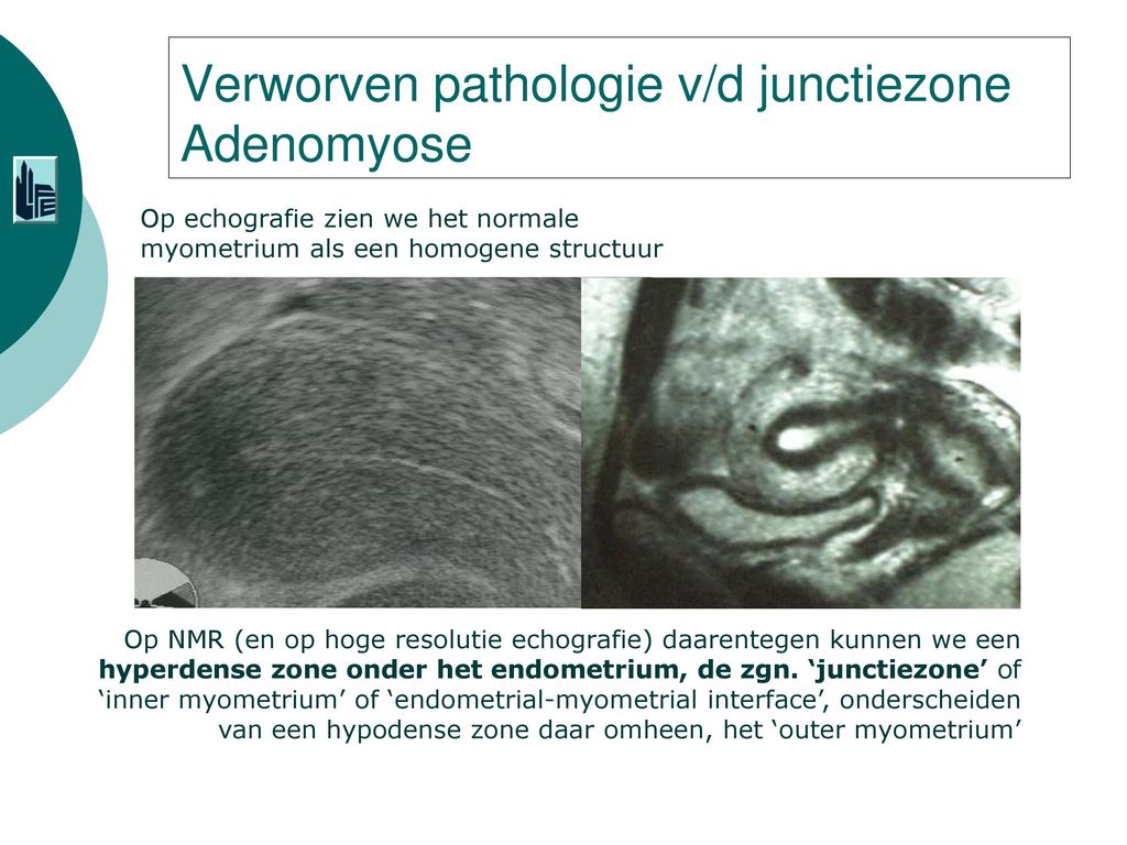
Criteria for adenomyosis on sonography were a globular uterus or asymmetric thickening of the anterior or posterior uterine wall, a poorly defined endometrial‐myometrial junction, a focal or diffuse heterogeneous myometrial echo texture, linear striated shadowing, myometrial cysts, and poorly defined foci with an abnormal myometrial echo texture without a discrete mass to suggest a myoma.
Figure 4.anechoic cystic lacunae in the posterior uterine wall (arrow) with a heterogeneous echo texture. Adenomyosis is a condition that many women haven't heard of, but it's way more common that you might expect. Criteria for adenomyosis on sonography were a globular uterus or asymmetric thickening of the anterior or posterior uterine wall, a poorly defined endometrial‐myometrial junction, a focal or diffuse heterogeneous myometrial echo texture, linear striated shadowing, myometrial cysts, and poorly defined foci with an abnormal myometrial echo texture without a discrete mass to suggest a myoma. The condition is typically found in women in the ages between 35 and 50. Adenomyosis is a condition that involves the encroachment, or movement, of the endometrial tissue that lines the uterus into the muscles of the uterus. Adenomyosis is a medical condition characterized by the presence of ectopic endometrial tissue (the inner lining of the uterus) within the myometrium (the thick, muscular layer of the uterus). As well as being misplaced in patients with this condition, endometrial tissue is completely functional. Linear striations (arrows) in the presence of a heterogeneous echo texture. In fact, a pretty substantial number of people go about their daily lives without. The strength and intensity of these changes as the condition develops. An enlarged uterus and painful, heavy periods can result. The adenomymosis advice association launched the first global digital survey in 2017, the results of which have been published on the youtube channel and distributed to the association's 400 clinicians worldwide. Anthony pompliano recommended for you
Criteria for adenomyosis on sonography were a globular uterus or asymmetric thickening of the anterior or posterior uterine wall, a poorly defined endometrial‐myometrial junction, a focal or diffuse heterogeneous myometrial echo texture, linear striated shadowing, myometrial cysts, and poorly defined foci with an abnormal myometrial echo texture without a discrete mass to suggest a myoma adenomyose. Adenomyomatosis of the gallbladder is a hyperplastic cholecystosis of the gallbladder wall.
Share :
Post a Comment
for "Adenomyose Echo : Fmpmc Ps Gynecologie Niveau Dcem2"

Post a Comment for "Adenomyose Echo : Fmpmc Ps Gynecologie Niveau Dcem2"